Table of Contents
3D Mammography (Tomosynthesis) vs 2D Mammogram: Which One Do You Need?

By Dr. Gajendra Kumar Himanshu


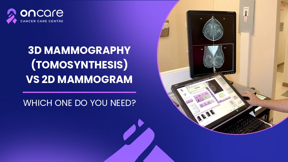
A 2D mammogram creates standard X-ray images of the breast, while 3D mammography (digital breast tomosynthesis) takes multiple images from different angles to create thin-slice views of breast tissue. Both are effective breast cancer screening methods, but 3D mammography may give doctors a clearer view of overlapping breast tissue and can be especially useful for some women with dense breasts.
In this blog, we will explain the difference between 2D and 3D mammograms, how each test works, their benefits and limitations, and when your doctor may suggest one over the other. The right choice depends on your age, breast density, symptoms, risk factors, previous mammogram results, and the advice of your doctor.
What Is a 2D Mammogram?
A 2D mammogram is the traditional form of mammography. It uses low-dose X-rays to create flat, two-dimensional images of the breast. Usually, images are taken from different views so that a radiologist can look for changes such as lumps, masses, or small calcium deposits called calcifications.
2D mammography has been used for many years and remains an important tool for breast cancer screening. It is widely available and can be used for both routine screening and evaluation of certain breast symptoms.
If you want to understand the warning signs that should not be ignored, you can also read our blog on breast cancer symptoms.
What Is 3D Mammography (Tomosynthesis)?
3D mammography, also called digital breast tomosynthesis (DBT), uses a similar X-ray process but takes several images while the machine moves in a small arc around the breast. A computer then combines these images into thin slices that allow the radiologist to examine the breast tissue layer by layer.
This can make some areas easier to see, especially when breast tissue overlaps. The experience is quite similar to a regular mammogram because the breast still needs to be compressed during the test.
3D mammography is often performed along with traditional or reconstructed 2D images.
2D vs 3D Mammogram: Key Differences
The main difference is how the images are created and viewed.
Feature | 2D Mammogram | 3D Mammography |
|---|---|---|
Image type | Flat, 2D images | Multiple thin-slice images |
Technology | Digital X-ray | Digital breast tomosynthesis |
View of overlapping tissue | Can be harder to interpret | Can separate tissue layers better |
Dense breasts | May be more difficult to read | May provide additional useful detail |
Screening use | Commonly used | Also used for screening |
Availability | Widely available | May not be available everywhere |
Cost | Usually lower | Often costs more |
Procedure | Breast compression required | Breast compression required |
Both methods can be used for breast cancer screening. Current evidence does not mean that every woman needs 3D mammography instead of 2D mammography.
What Are the Benefits of 3D Mammography?
One reason doctors may suggest 3D mammography is that it can provide more detailed views of breast tissue.
Better view of overlapping tissue
In a traditional 2D image, normal breast tissue can overlap and sometimes make an abnormal area harder to see. 3D mammography provides thin images through the breast, which may help the radiologist examine these areas more clearly.
Helpful for some women with dense breasts
Women with dense breasts have more fibrous and glandular tissue compared with fatty tissue. This can make mammograms more difficult to interpret. Studies suggest that 3D mammography can be helpful in some women with dense breasts.
May reduce callbacks
Some studies have found that 3D mammography can reduce the chance of being called back for additional imaging after screening. However, results can vary between women and screening settings.
Are There Any Limitations of 3D Mammography?
3D mammography is not automatically the right choice for everyone.
The first consideration is cost and availability. 3D mammograms can cost more than standard 2D mammograms, and coverage may differ depending on your insurance or healthcare plan.
Like 2D mammography, 3D mammography also uses X-rays and requires breast compression. Some women may experience temporary discomfort during the procedure.
Also, more detailed images do not mean that every cancer will be found. Mammography has limitations, and some women may need additional tests such as ultrasound or MRI based on their individual situation.
Which One Do You Need?
There is no single answer for every woman.
Your doctor may consider:
- Your age and overall breast cancer risk
- Whether you have dense breast tissue
- Any breast symptoms or recent changes
- Your previous mammogram results
- Family history of breast cancer
- Previous breast procedures or biopsies
- The availability and cost of 3D mammography
Both 2D digital mammography and 3D mammography are considered effective screening options. Screening mammography is generally recommended every two years for women aged 40 to 74 at average risk, while screening decisions for people outside that group may need to be individualised.
If you have a breast lump, nipple discharge, skin changes, or another new breast concern, do not wait for your routine screening. Talk to a doctor about whether diagnostic imaging is needed.
3D vs 2D Mammogram: Is 3D Always Better?
Not necessarily. 3D mammography offers a different and sometimes more detailed way of viewing breast tissue, but it does not mean that every woman must have a 3D scan.
Research has found some benefits with 3D mammography, including lower recall rates in some studies and improved detection of certain cancers. At the same time, evidence does not show a simple one-size-fits-all advantage for every screening situation.
The most suitable test should be decided according to your individual risk and your doctor’s recommendation.
If you want to learn more about mammograms, then you can visit the official website of the National Cancer Institute.
Consult Today
If you are unsure whether you need a 2D mammogram, 3D mammography, ultrasound, or another breast imaging test, speaking with a specialist can help you understand your options.
At Oncare Cancer Hospital, the cancer care team can review your symptoms, medical history and previous reports and guide you toward the appropriate next step. Early evaluation of a breast change can help avoid unnecessary delays in diagnosis and treatment.
Disclaimer
This information is for educational purposes only and does not constitute medical advice, diagnosis, or treatment. Always discuss decisions about breast screening, imaging tests, or any cancer treatment with a certified cancer healthcare provider.





Frequently Asked Questions
3D mammography can provide more detailed views of breast tissue and may be helpful for some women, including some women with dense breasts. However, both are effective screening methods.
The experience is similar to a standard mammogram. Breast compression is required, which may cause temporary discomfort.
It may be useful for some women with dense breast tissue or certain screening needs. Your doctor can decide whether it is appropriate based on your individual situation.
3D mammography is commonly performed with traditional or reconstructed 2D images. The imaging approach used depends on the equipment available and the clinical situation.
Written and Verified by:
Dr. Gajendra Kumar Himanshu Exp: 10 Yr
Medical Officer
Book an Appointment
Related Blogs

Esophagectomy: Food Pipe Cancer Surgery and How to Eat After
Learn about esophagectomy for food pipe cancer, including the surgery, recovery, dumping syndrome, nutrition, and follow-up at Oncare Cancer Hospital.

Superior Vena Cava Syndrome: A Cancer Emergency Causing Face and Neck Swelling
Learn about superior vena cava syndrome, its symptoms, diagnosis, emergency warning signs, stent placement, and treatment options at Oncare Cancer Hospital.

Neutropenic Diet: Safe Eating When Your Immunity Is Low During Chemotherapy
Learn about the neutropenic diet during chemotherapy, including safe foods, food safety, nutrition, and infection prevention at Oncare Cancer Hospital.