Table of Contents
Immunohistochemistry (IHC) Testing: How Labs Identify Your Exact Cancer Type

By Dr. Gajendra Kumar Himanshu


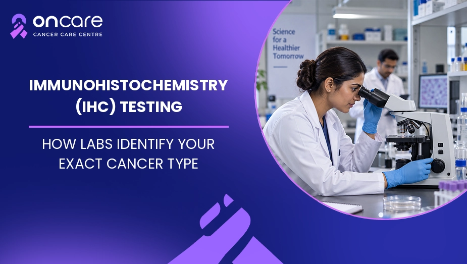
Immunohistochemistry (IHC) is a laboratory test that uses special antibodies to detect specific proteins, called markers, in a tissue sample. By seeing which markers are present or absent, a pathologist can help identify the type and subtype of cancer and sometimes determine where a cancer may have started. IHC can also provide information that helps doctors choose certain treatments.
When a biopsy shows abnormal or cancerous cells, seeing them under a microscope may not be enough to identify the exact type of cancer. This is where IHC can help.
In this blog, we will explain what IHC testing is, why doctors recommend it, how the test is performed, what common markers mean, how IHC can influence treatment, and how to understand the results in simple words.
What Is Immunohistochemistry (IHC) Testing?
Immunohistochemistry, commonly called IHC, is a laboratory method used to find specific proteins or other markers in tissue. It uses antibodies that attach to particular markers inside or on the surface of cells. The reaction creates a visible stain that a pathologist can examine under a microscope.
Think of IHC as a way of giving the pathologist extra clues about a group of abnormal cells. The shape and appearance of the cells provide the first clues. IHC adds more information by showing which proteins those cells carry.
IHC is usually performed on tissue obtained through a biopsy or surgery. If you want to understand the first step of getting a cancer diagnosis, you can read this blog about biopsy tests.
Why Is IHC Needed in Cancer Diagnosis?
A pathologist first examines a tissue sample under a microscope. In some cases, the appearance of the cells is enough to identify the cancer. In other cases, different cancers can look similar.
IHC can help distinguish between these possibilities. It may help determine whether a tumor is a particular type of carcinoma, lymphoma, melanoma, sarcoma, or another cancer. It can also help identify the likely site where a cancer began, especially when cancer is found in a location where its origin is not obvious.
For example, if cancer cells are found in a lymph node, the doctor may need to know whether they started in that lymph node or spread there from another organ. A carefully selected group of IHC markers can provide useful clues.
You can also learn more about how cancer is diagnosed and why doctors may use several tests before confirming a diagnosis.
How Does IHC Testing Work?
IHC testing is performed on a tissue sample that has usually already been collected during a biopsy or surgery.
Step 1: Tissue Is Collected
A doctor removes a small sample of the suspicious tissue during a biopsy. In some cases, tissue removed during surgery is used for testing.
Step 2: The Tissue Is Prepared
The tissue is preserved and processed in the pathology laboratory. Very thin sections are placed on glass slides.
Step 3: Special Antibodies Are Added
The laboratory adds antibodies designed to attach to specific markers in the cells. Each antibody looks for a particular protein or marker.
Step 4: The Marker Becomes Visible
When an antibody attaches to its target, a chemical or dye-based reaction produces a visible stain. The pathologist then examines the stained tissue under a microscope.
Step 5: The Results Are Interpreted
The pathologist looks at the staining pattern, how strong the staining is, and which cells are stained. These findings are combined with the appearance of the tissue, the patient's clinical information, scans, and other laboratory tests.
This means that IHC is not usually interpreted as a single yes-or-no test. The overall pattern of several markers is often more useful than one marker by itself.
What Can IHC Tell Doctors About Cancer?
IHC can answer different questions depending on the cancer and the markers selected.
It may help doctors:
- Identify the type or subtype of a tumor.
- Find clues about where a cancer started.
- Distinguish cancer from some noncancerous conditions.
- Identify certain proteins that may affect treatment.
- Provide additional information when the diagnosis is difficult.
IHC is particularly useful when cancer cells have an unusual appearance or when the cancer's original site is uncertain.
Common IHC Markers Used in Cancer
There are many different IHC markers, and the combination used depends on the suspected cancer. A marker is not automatically a sign of cancer. It is a protein or other feature that can provide information about the cells.
IHC marker or group | Where it may be useful | What it can help doctors understand |
|---|---|---|
ER | Breast cancer | Whether cancer cells have estrogen receptors |
PR | Breast cancer | Whether cancer cells have progesterone receptors |
HER2 | Breast and some other cancers | Whether HER2 protein is overexpressed |
Ki-67 | Several cancers | Information about how actively cells are dividing |
TTF-1 | Mainly lung and thyroid tumors | Can provide clues about tumor origin |
CD markers | Blood cancers and some other tumors | Help identify certain cell types |
PAX8 | Some kidney, thyroid, and gynecologic tumors | Can provide clues about tissue of origin |
The exact markers used are selected by the pathologist based on the tissue appearance and the clinical question. Not every patient needs every marker.
Why ER, PR and HER2 Matter in Breast Cancer
For example, in breast cancer, ER, PR, and HER2 results can provide important information for treatment planning. The National Cancer Institute notes that these biomarker results may help identify treatments that are appropriate for an individual patient.
Can IHC Help Find Where Cancer Started?
Yes, one important use of IHC is helping doctors identify the likely site of origin of a cancer.
Sometimes a patient has metastatic cancer, meaning cancer has spread from one part of the body to another. For example, cancer may be found in a lymph node, liver, bone, or lung, while the original tumor is not immediately clear.
The pathologist may use a panel of IHC markers to compare the tumor's pattern with patterns commonly seen in different cancers. This can help narrow down the possible origin.
However, IHC does not always identify the original cancer with certainty. Some tumors share the same markers, so doctors may need imaging, clinical history, molecular testing, or other investigations.
How Does IHC Help With Cancer Treatment?
IHC is not only about naming the cancer. Some IHC markers can also provide information about whether a particular treatment may be useful.
For example, HER2 testing can help identify cancers with HER2 protein overexpression. In breast cancer, estrogen and progesterone receptor testing can help doctors decide whether hormone therapy may be appropriate.
Some other IHC-based biomarkers can also help guide treatment decisions in selected cancers. The College of American Pathologists notes that validated IHC assays can be important for predictive markers that guide cancer treatment.
If you are exploring treatment after a cancer diagnosis, you can read more about cancer treatment options and discuss your specific pathology results with your oncologist.
What Does an IHC Result Look Like?
An IHC report may list the name of each marker tested and describe whether the result is positive, negative, weak, moderate, or strong. Some tests may also provide a percentage or score.
For example, a report may say that a particular marker is positive or negative. However, the meaning depends on the marker, cancer type, testing method, and clinical situation.
A positive result does not always mean that a person has a particular cancer. Similarly, a negative result does not always completely rule out a cancer. Pathologists interpret IHC results as part of the complete pathology assessment.
This is why patients should avoid trying to interpret one IHC result without looking at the full pathology report and medical history.
Is IHC the Same as Genetic Testing?
No, IHC and genetic or molecular testing look at different features of cancer.
IHC mainly looks for proteins or other tissue markers, while molecular tests may look for changes in genes, chromosomes, or other molecular features of cancer cells.
Sometimes both types of testing are needed. If IHC does not provide a clear answer, doctors may recommend molecular testing or another laboratory test.
Modern cancer diagnosis may combine microscopy, IHC, imaging, and molecular tests to create a clearer picture of the tumor.
If you want to read more about immunohistochemistry (IHC), you can visit the National Cancer Institute.
Consult Today
Getting an accurate cancer diagnosis is an important first step because treatment depends on the type and features of the cancer. IHC testing can give pathologists important information about the proteins present in tumor cells and can help distinguish between cancer types, identify possible tumor origin, and, in some cases, guide treatment decisions.
At Oncare Cancer Hospital, patients can discuss their biopsy, pathology, IHC results, and treatment options with cancer specialists. If your doctor has recommended IHC testing or you have received an IHC report that you do not understand, consult your cancer care team before making decisions based on individual test results.
Disclaimer
This information is for educational purposes only and does not constitute medical advice, diagnosis, or treatment. IHC results must be interpreted along with the complete pathology report and each patient's clinical details. Always discuss your test results and treatment decisions with a certified cancer healthcare provider.





Frequently Asked Questions
IHC is a laboratory test that uses antibodies to detect specific proteins or markers in tissue. It helps pathologists identify and classify certain cancers.
Not necessarily. An IHC result must be interpreted along with the tissue appearance, other tests, and the patient's clinical information.
Yes, IHC can help distinguish between different types or subtypes of cancer and may provide clues about where a tumor started.
Yes, certain IHC biomarkers, such as ER, PR, and HER2 in appropriate cancers, can provide information that helps doctors select treatment.
Written and Verified by:
Dr. Gajendra Kumar Himanshu Exp: 10 Yr
Medical Officer
Book an Appointment
Related Blogs

Frozen Section Biopsy: How Doctors Take Decisions During Surgery
Learn what a frozen section biopsy is, how it is done during surgery, and its uses, results, benefits, and limitations with guidance from Oncare Cancer Hospital.
Pleural Tap (Thoracentesis): Draining Fluid Around the Lungs in Cancer
Learn what a pleural tap is, why it is done in cancer, how it helps with breathing, its risks, and what happens if fluid returns. Oncare Cancer Hospital's guidance.

Chemotherapy-Induced Peripheral Neuropathy: Managing Numbness and Tingling
Learn about chemotherapy-induced neuropathy, including numbness, nerve pain, and weakness. Know simple ways to manage symptoms with Oncare Cancer Hospital.